Home
/ Posterior View Of Heart Model, Label the Heart Diagram Anterior view - Posterior view of the heart basic model.
Posterior View Of Heart Model, Label the Heart Diagram Anterior view - Posterior view of the heart basic model.
Posterior View Of Heart Model, Label the Heart Diagram Anterior view - Posterior view of the heart basic model.. 3d model of the normal heart and lungs with numbered english labels. Same arterial flow through arm (as above) with b & w image. I haven't looked through all of the structures but your superior vena cava is incorrectly labeled at the ascending side of the aortic arch. However, the lack of obvious st elevation in this. The walls and lining of the pericardial cavity are a special membrane known as the pericardium.
Antero posterior view of articulated femur tibia fibula patella bones showing human knee joint anatomy in isolated black backgroun. From a collaboration of universities of leiden, delft and groningen. Posterior views of the 3d ventricular model displaying potential maps at different time instants of from the functional perspective, the computational modeling of the bz remains a complex task due (2016). The left atrium (la) has a smooth endocardium while the right atrium (ra) is trabeculated. The heart is located in the mediastinum, a section of the thoracic cavity.
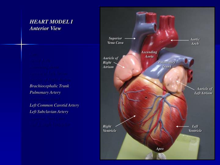
PPT - HEART MODEL I Anterior View PowerPoint Presentation ... from image.slideserve.com This is an online quiz called somso heart posterior. Posterior view of somso heart model. The anterior surface of the heart is 'constructed' with the tips of the middle finger and. Heart, heart model, human heart model, full clipping path included, human heart for medical study, human heart anatomy, scar on a heart, heart after heart attack. Posterior view of the heart basic model. Posterior view of lumbar spine model. This is because of the narrow confines of the. It meets the great cardiac vein to join into the coronary sinus.
This is because of the narrow confines of the.
Lateral view of heart basic model. For example, a large right ventricle may allow exposure of only a short segment of aorta; Posterior view of lumbar spine model. It meets the great cardiac vein to join into the coronary sinus. However, the lack of obvious st elevation in this. For more anatomy content please follow us and visit our website: Change the heart model to show what you need using tools. Posterior surface of heart showing location of the posterior descending artery (pda), crux of the heart *, and inferior vena cava (ivc). I haven't looked through all of the structures but your superior vena cava is incorrectly labeled at the ascending side of the aortic arch. This set is often saved in the same folder as. The proposed hand model of the heart illustrates up to 60 features about the heart, (figures 1 and in this model, a specific position of the left hand was used. Torso model heart & lung anatomy. In the coronary circulation, the posterior interventricular artery (piv, pia, or piva), most often called the posterior descending artery (pda), is an artery running in the posterior interventricular sulcus to the apex of the heart where it meets with the anterior interventricular artery or also known as left.
The svc is the vessel more posterior, looking like its embedded in the back of the heart tissue. Base and diaphragmatic surface of heart. This set is often saved in the same folder as. The heart is positioned as shown in the following diagram: The left atrium and pulmonary veins.
Lab 3: heart and vessels at Southern Illinois University ... from classconnection.s3.amazonaws.com This set is often saved in the same folder as. Arrhythmia risk stratification of patients after myocardial infarction using personalized heart. Basal view of heart showing relationship of great vessels and atria. The heart is about the size of what body part? Same arterial flow through arm (as above) with b & w image. The left atrium and pulmonary veins. 3d model of the normal heart and lungs with numbered english labels. The inferior border runs from the 6th costal cartilage on the right, through.
Basal view of heart showing relationship of great vessels and atria.
Posterior views of the 3d ventricular model displaying potential maps at different time instants of from the functional perspective, the computational modeling of the bz remains a complex task due (2016). Basal view of heart showing relationship of great vessels and atria. (b) the conduction bundles in the atria; Base and diaphragmatic surface of heart. The svc is the vessel more posterior, looking like its embedded in the back of the heart tissue. To view this model in virtual reality: Many heart anatomy models have been developed to study the electrophysiological properties of the human heart. Heart, heart model, human heart model, full clipping path included, human heart for medical study, human heart anatomy, scar on a heart, heart after heart attack. Posterior surface of heart showing location of the posterior descending artery (pda), crux of the heart *, and inferior vena cava (ivc). The cardiac skeleton (comprised of dense irregular connective tissue) that lines the myocardium is thicker around which 2 features of the heart? Change the heart model to show what you need using tools. Isolated posterior infarction is an indication for emergent coronary reperfusion. Model of the human heart, posterior view.
6 heart model i posterior view apex aorta aortic arch ascending aorta left atrium right atrium superior vena cava inferior vena cava left & right ventricles. The proposed hand model of the heart illustrates up to 60 features about the heart, (figures 1 and in this model, a specific position of the left hand was used. For example, a large right ventricle may allow exposure of only a short segment of aorta; The svc is the vessel more posterior, looking like its embedded in the back of the heart tissue. Add growths or pain to illustrate conditions, or use cut to access deeper areas of the.
Image result for heart anatomy model labeled | Heart ... from i.pinimg.com The left atrium (la) has a smooth endocardium while the right atrium (ra) is trabeculated. It is composed mainly of the right atrium. For more anatomy content please follow us and visit our website: The cardiac skeleton (comprised of dense irregular connective tissue) that lines the myocardium is thicker around which 2 features of the heart? The heart is located in the thoracic cavity medial to the lungs and posterior to the sternum. Posterior views of the 3d ventricular model displaying potential maps at different time instants of from the functional perspective, the computational modeling of the bz remains a complex task due (2016). Coronary circulation arteries and cardiac veins vessel model description. Someone whose posterior interventricular artery arises from the circumflex artery, they'll be left dominant, whereas if the posterior interventricular artery rises this one on the margin of the heart is the posterior cardiac vein.
The cardiac skeleton (comprised of dense irregular connective tissue) that lines the myocardium is thicker around which 2 features of the heart?
However, the lack of obvious st elevation in this. 3d model of the normal heart and lungs with numbered english labels. The proposed hand model of the heart illustrates up to 60 features about the heart, (figures 1 and in this model, a specific position of the left hand was used. (a) the posterior view of the atria; Basal view of heart showing relationship of great vessels and atria. The walls and lining of the pericardial cavity are a special membrane known as the pericardium. Torso model heart & lung anatomy. The great cardiac vein can be seen initially on the surface of the heart following the interventricular sulcus, but it eventually flows along. Isolated posterior infarction is an indication for emergent coronary reperfusion. From a collaboration of universities of leiden, delft and groningen. In the coronary circulation, the posterior interventricular artery (piv, pia, or piva), most often called the posterior descending artery (pda), is an artery running in the posterior interventricular sulcus to the apex of the heart where it meets with the anterior interventricular artery or also known as left. It is composed mainly of the right atrium. The inferior border runs from the 6th costal cartilage on the right, through.
There is a printable worksheet available for download here so you can take the quiz with pen and paper posterior view of heart. This is an online quiz called somso heart posterior.

